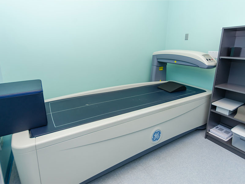
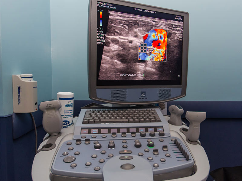
Diagnostic imaging
The collection of data is volumetric allowing a non-invasive endoscopic view, three-dimensional reconstruction, images of preventive diagnosis like: Angiography CT, Pulmonary analysis CT, among others.
The speed with which the information is acquired allows for pediatric or geriatric studies, avoiding sedation in many cases .It also optimizes the use of intravenous contrast, with the synchronized use of the automatic injector, works with the maximum peaking of intensity and thus the dosage is minimum.
- Capacity for automatic programming
- Speedy acquisition
- Synchronized studies with cardiac and/or respiratory monitoring
- Application in skeletal, thoracic-abdominal-pelvic and neurovascular muscles
- Multi-element antennas cover more anatomy without jeopardizing quality
The acquisition of this technology optimizes the use of radiation, imaging plate (IP) screens in response to an increase in radiation sensibility, taking note of the interest in radiological practice to protect all personnel, exposed to radiation through their work, the public and the environment.
The CR system of AGFA harmonizes with the environment by eliminating the use of processing chemicals.
- Given the sensibility of the recipient the dose of radiation is reduced
- Greater speed in the subprocess of the diagnostic imaging system
- Increase in information upon the application of software packages for digital manipulation, thus supplementing diagnostic tools
- Easy to store, share and ship long distance, thus promoting referrals
- Easily reproduced and taken away by the patient in a pleasant way
This is the standard established form to measure bone mineral density and is a rapid and painless procedure to measure the loss of bone tissue.
- High acquisition speed
- Easy image processing
- Cinematic viewing
- A similar display to CAT and MR
- Updating service available
Ultrasound, also known as sonography or echography, is a diagnostic procedure which transmits sound waves of high frequency, inaudible to the human ear, through the tissues of the body. The echoes are registered and transformed into video or photographic images of the internal structures of the body. Ultrasound images help in the diagnosis of a wide range of illnesses and conditions. Ultrasound is used to create images of soft tissue structures, for vascular studies, of joints and small parts like thyroids, testicles, eyes, etc. Ultrasound can also be used with other diagnostic procedures like a directed biopsy, drainage of liquid collection and abscesses.
The Clínica Bíblica Hospital has the latest technology in ultrasound equipment, like equipment of the Fourth Dimension for obstetric studies in general, and technological equipment like HD Life, which has high resolution transducers, capable of making evaluations with the best resolution on the market.
Image gallery
https://www.clinicabiblica.com/en/services/diagnostic-imaging#sigFreeId823b712ca2
We believe in the technology and innovation that saves lives.
For this reason we have acquired the first Digital Mammography with Microdose technology in Central America.
It is recommended that women have a mammogram once a year, after 40 years of age. Breast tissue is sensitive to radiation, so that it is important that the dose be as low as possible.
Microdose technology by Philips makes it possible to reduce to one half the dose of radiation that the patients receive when having a mammography exam.
Image Gallery
https://www.clinicabiblica.com/en/services/diagnostic-imaging#sigFreeId0bc142c363
Our location
We are located in the Cabezas López building, main floor. Consult the map for our location.
Telephone Exchange: (506) 2522-1000.
Map
https://www.clinicabiblica.com/en/services/diagnostic-imaging#sigFreeId7fafdf36d6